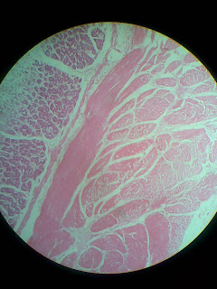
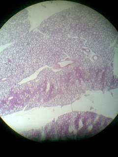
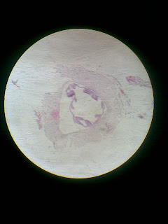
More than 100 Slides for Histology lab. click
 |
| Colon and rectum...jpg |
 |
| Breasts..lactating phase 23.jpg |
 |
| ADRENAL~1.JPG |
 |
| ADRENAL~1.JPG |
 |
| Adrenal8.jpg |
 |
| Adrenal9.jpg |
 |
| Appendix.8.jpg |
 |
| Appendix15.jpg |
 |
| Appendix..46.jpg |
 |
| Appendix..5+¦7.jpg |
 |
| Appendix..84.jpg |
 |
| Appendix..85.jpg |
 |
| Appendix..debry present in lumen..intestinal glands less in number.jpg |
 |
| Appendix..lymphocytes present.debry in lumen.jpg |
 |
| Breasts..lactating phase 23.jpg |
 |
| Cervix and vagina.jpg |
 |
| Colon and rectum...jpg |
 |
| Colon and rectum..2.jpg |
 |
| Cornea........jpg |
 |
| Cornea......jpg |
 |
| Cornea....jpg |
 |
| Duod..jpg |
 |
| Duod.2.jpg |
 |
| Duod.2..LEAf like villi and brunner's glands.jpg |
 |
| Duodenum.+¿3+¦.jpg |
 |
| Duodenum..23.jpg |
 |
| Duodenum..25.jpg |
 |
| Ear.americ+ñn.jpg |
 |
| Ear.american.jpg |
 |
| Epididymis and testes..65.jpg |
 |
| Epididymis and testes.jpg |
 |
| Epididymis.aj...jpg |
 |
| Fallopian tube and broad ligament..jpg |
 |
| Fallopian tube..58.jpg |
 |
| Fallopian tube..mucosal lining thrown into longitudinal folds..it is continuous at upper left part of this pic with broad ligament..jpg |
 |
| GALLBL~1.JPG |
 |
| Gall bladder.28.jpg |
 |
| Ileum+¿..CLUB SHAPED VILLI AND PEYERS PATCHES.jpg |
 |
| Ileum...8.jpg |
 |
| Ileum...jpg |
 |
| Ileum..OUTER.jpg |
 |
| Ileum2.jpg |
 |
| Duodenum..23.jpg |
 |
| Duodenum..25.jpg |
 |
| Jujenum.ELONGATED VILLI.no peyers patches.no submucosal gland.jpg |
 |
| Kidney...PCT AND DCT are present..jpg |
 |
| Kidney.medula is present..collecting ducts seen.jpg |
 |
| Liver..6pg7.jpg |
 |
| Liver58.jpg |
 |
| Lungs..5.jpg |
 |
| Lungs.98.jpg |
 |
| Oesophagus..+¦3.jpg |
 |
| Oesophagus..st.squmamous epithelium in upper part.columnar in lower part..inner circular outer longitudinal.mixed glands.jpg |
 |
| Oesophagus..65.jpg |
 |
| Ovary..5.jpg |
 |
| Ovary..62.jpg |
 |
| Pancreas+¿.jpg |
 |
| Pancreas..jpg |
 |
| Pancreas..outer.jpg |
 |
| Pancreas.adipocytes are seen.jpg |
 |
| Pancreas.in the center interlobular duct is present.red colored islets of langerhans are present..in addition.adipocytes are also seen.jpg |
 |
| Pancreas.jpg |
 |
| Pancreas.pale colored islets of langerhans present.jpg |
 |
| Pancreas2.jpg |
 |
| Pancreas32.jpg |
 |
| Parotid gland+¦8.jpg |
 |
| Parotid gland92.jpg |
 |
| Parotid gland..8.jpg |
 |
| Pituitary..2.jpg |
 |
| Pituitary..anterior pituitary.darkly staining chromophil cells and lightly staining chromphobe cells are present...jpg |
 |
| Pituitary..hering bodies are seen as light redded spots in the posterior pituitary..jpg |
 |
| Prostate...6+¼.jpg |
 |
| Prostate..6.jpg |
 |
| Prostate..sadaken.jpg |
 |
| Seminal vesicle...64.jpg |
 |
| Seminal vesicle.89.jpg |
 |
| Stomach 50.jpg |
 |
| Stomach...jpg |
 |
| Stomach..3.jpg |
 |
| Stomach..40.jpg |
 |
| Sublingual gland...jpg |
 |
| Submandibular gland..3+¦.jpg |
 |
| Submandibular..95.jpg |
 |
| Thyroid gland..3.jpg |
 |
| Tongue..23.jpg |
 |
| Tongue..34.jpg |
 |
| Tongue..38.jpg |
 |
| Tongue.fungiform papillae.jpg |
 |
| Trachea...3+¦.jpg |
 |
| Trachea...jpg |
 |
| Trachea..OA.jpg |
 |
| Trachea..hyaline cartilage is seen as a c shaped cartilage...jpg |
 |
| Trachea..hyaline cartilage...jpg |
 |
| Trachea..k+¦.jpg |
 |
| Ureter...jpg |
 |
| Urinary bladder 50.jpg |
 |
| Urinary bladder..84.jpg |
 |
| Uterus6k.jpg |
 |
| Uterus..BLUe overall.jpg |
 |
| Uterus...6.jpg |
 |
| Vas deferens..outer and inner longitudinal middle circular layers are present..epithelium is pseudostratified columnar epithelium.jpg |
 |
| Vas defferens...851.jpg |
No comments:
Post a Comment